Pathology has been intricately linked to Singapore's history and healthcare since its colonial days. The history of pathology began with the arrival of the late Dr George Alexander Finlayson in Singapore on 12 May 1903, when he took up the appointment of Municipal Bacteriologist.1 118 years later, we co-celebrate #pathology118 with Singapore General Hospital's bicentennial birthday.
In my job as a specialist anatomical pathologist, I am blessed to be able to marvel at the beauty of pathology day in, day out. In this photo essay, I hope to provide a sneak peek into that natural beauty, so come along for a visual stroll through the garden of pathology.
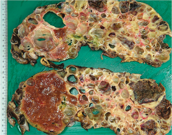
Undoubtedly, the macroscopic manifestation of disease provokes the most visceral reaction (pun intended) to pathological beauty. The above photograph is a striking example of a polycystic liver, with innumerable variably sized cysts scattered throughout the liver parenchyma.
Armed with stains for every occasion
The haematoxylin and eosin (H&E) stain is the most widely used histochemical stain in histopathology and it is a combination of two stains: haematoxylin, which stains cell nuclei purple, and eosin, which stains the cytoplasm and extracellular matrix pink. In this example of a hepatocellular carcinoma, the H&E stain perfectly demonstrates the contrast between the abundant pink cytoplasm and round purple nuclei of the hepatocytic tumour cells.

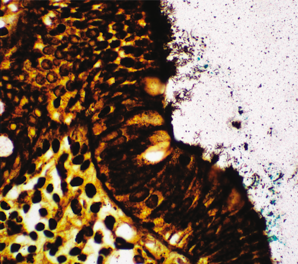
Analogous to the various photo filters in our smartphones, the Histopathology Laboratory has an arsenal of special histochemical stains that allows us to accentuate various pathologies. This Warthin-Starry histochemical stain highlights the spirochetes embedded along the luminal borders of intestinal epithelial cells.
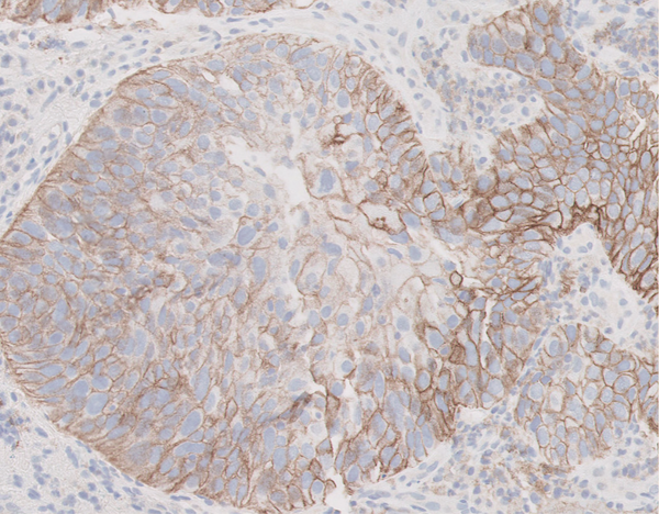
Immunohistochemistry (IHC) allows for the identification of specific tissue proteins via the binding of specifically raised antibodies and an accompanying chromogenic reaction. This revolutionary technique allows us to continue providing accurate diagnoses, discover new diagnostic entities and even provide therapeutic information. The oesophageal squamous carcinoma cells express programmed death-ligand 1 (an immune checkpoint protein) in a positive membranous staining pattern, predicting that the patient is likely to respond to immuno-oncotherapy.
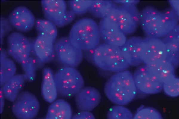
Photo: Clinical A/Prof Alvin Lim Soon Tiong and Ms Lim Tse Hui, Singapore General Hospital Cytogenetics Laboratory, Department of Molecular Pathology
In instances where the interpretation of IHC is equivocal, we turn to our pathology colleagues at the Cytogenetics Laboratory for help. This fluorescence in situ hybridisation image shows amplification of the human epidermal growth factor receptor 2 (HER2) gene (in red) in breast cancer tumour cells. Observe the relatively increased HER2 signals compared to the green control probe targeting centromere 17.
Cutting-edge technology
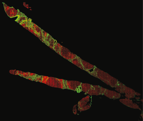
Photo: Dr Dean Tai and Mr Ren Yayun, HistoIndex Pte Ltd
In my academic research collaborations, I have been fortunate to experience novel technologies that allow us to admire pathologies with higher clarity and definition. These two liver tissue core biopsies are imaged with second harmonic generation microscopy, a tissue imaging system that uses nonlinear optical microscopy to observe endogenous tissue signals in unstained tissue samples.2 Look at the bright green fibrous bands in this cirrhotic liver.
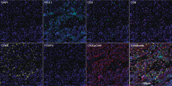
Photo: Dr Joe Yeong and Ms Justina Lee, Integrative Biology for Theranostics Laboratory
Immuno-oncotherapy is a major breakthrough in the fight against cancer, and has heralded intense research into the tumour microenvironment. Quantitative multiplex immunofluorescence opens the door to spatial phenotyping, allowing pathologists to simultaneously look at multiple cell proteins in order to unravel potential biomarkers, all while using only one section of tissue! The composite image above shows the various immune cells (highlighted in cyan, green, magenta, yellow and orange) among the colorectal cancer cells (in red).
Artificial intelligence (AI) is permeating all aspects of life. In the pursuit of laboratory digitalisation, collaborations with medical technology start-ups will yield the development of pathology-focused and AI-driven applications that can assist busy pathologists in their diagnostic work. Wonder at the abilities of a deep-learning convolutional neural network as it detects malignant colonic glands in the whole slide image on the left.3
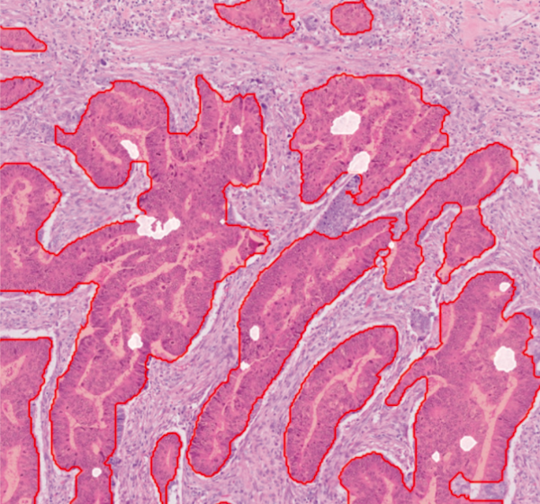
Photo: Dr Sahil Ajit Saraf, Qritive Pte Ltd
Thank you for taking a moment to walk with me through this garden of pathology. Like me, I hope you find the beauty of pathology mesmerising. As pathologists, and more importantly doctors, it is our fervent hope that by understanding the basis of diseases that lie behind every glass slide, we will be able to find a cure and alleviate the suffering of the patient.
